Fat necrosis management is crucial for those dealing with this condition involving damaged breast tissue and fatty tissue content. Understanding its causes, such as skin necrosis and fibrotic fat necrosis, and symptoms like a lump can help in seeking the right treatment for presentation fat necrosis. This blog post dives into effective strategies for managing fat necrosis, including lifestyle changes, medical interventions, and addressing the lump. We will also explore how to recognize the signs of breast fat necrosis and pancreatic fat necrosis early on, based on recent understanding from a necrosis article related to fat tissue necrosis.
With expert insights and practical tips, you’ll learn how to navigate this issue confidently. Whether you’re facing breast fat necrosis yourself or supporting someone who is, having the right information can make a difference. Let’s break down what you need to know to tackle fat necrosis head-on.
What is Fat Necrosis?
Definition
Fat necrosis refers to the death of fat tissue due to injury or trauma. This condition can occur when there is damage to the fat cells, leading them to die and form necrotic tissue.
Locations
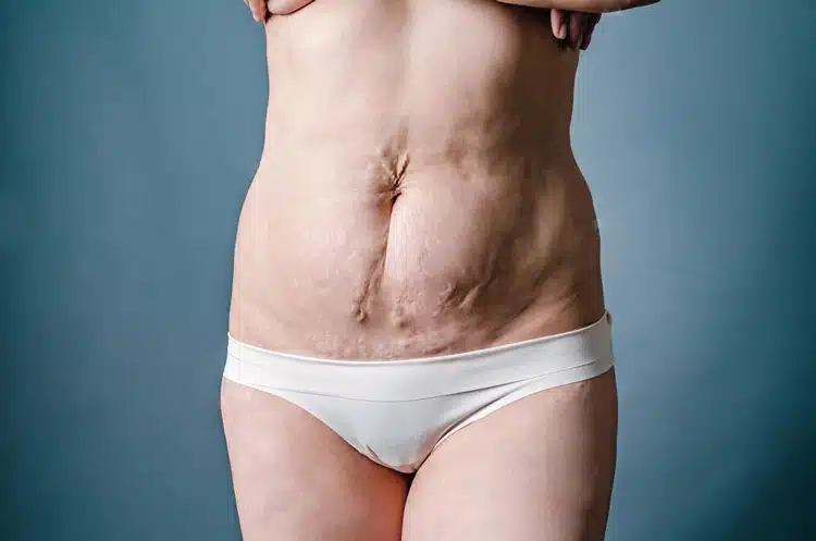
Fat necrosis can develop in various parts of the body. It most commonly occurs in the breast. However, it can also happen in areas such as the abdomen, around the pancreas, or in breast fat necrosis. Each location may present different symptoms.
Symptoms
The condition often results in lumps or nodules. These formations, such as breast fat necrosis, are typically benign, meaning they are not cancerous. In many cases, people may discover these lumps during a breast examination. The presence of these nodules, such as breast fat necrosis, can cause concern, but they usually do not require aggressive treatment.
Causes
Several factors can lead to fat tissue necrosis. Trauma is a common cause. For example, surgery or injury to the breast can result in this condition. Other causes include infections, breast fat necrosis, or conditions that affect blood flow to the area. Understanding these causes helps in managing fat necrosis effectively.
Diagnosis
Doctors use imaging techniques to diagnose fat necrosis. Mammograms and ultrasounds are common methods for examining breast lumps. These tools help distinguish between fat necrosis and other conditions, such as tumors. A biopsy may be necessary if there is uncertainty about the nature of the lump.
Management
Management of fat necrosis focuses on observation and reassurance. Most cases do not need surgical intervention. Regular monitoring through follow-up appointments is often sufficient. If symptoms worsen or if there are concerns about malignancy, further evaluation may be needed.
Recent Understanding
Recent studies have improved the understanding of fat necrosis. Medical professionals now recognize that it is a common occurrence after procedures like liposuction or breast surgery. Awareness of this condition has increased among healthcare providers and patients alike.
Emotional Impact
The diagnosis of fat necrosis can cause anxiety for patients. Many worry about potential complications or misdiagnosis as cancer. Education about the benign nature of this condition can help alleviate fears.
Causes of Fat Necrosis
Blunt Trauma
Blunt trauma is a significant cause of fat necrosis. This type of injury can occur from falls, car accidents, or sports injuries. The impact damages the fatty tissue. As a result, it leads to localized inflammation and cell death.
Surgical procedures can also result in fat necrosis. Operations that involve cutting through fatty tissue often lead to this condition. For example, surgeries on the breast can disrupt the surrounding fat. This disruption creates an environment for fat necrosis to develop.
Radiation Therapy
Radiation therapy contributes to fat necrosis as well. Patients undergoing treatment for cancer may experience damage to fatty tissues. The radiation can cause inflammation and necrosis in these areas. It often occurs in regions treated for breast cancer or other cancers near fatty tissue.
Studies show that this side effect is common among patients receiving radiation. The damaged cells can lead to complications long after treatment ends. Managing these effects is crucial for patient recovery.
Acute Pancreatitis
Severe acute pancreatitis is another factor linked to fat necrosis. This condition causes inflammation of the pancreas, leading to the release of digestive enzymes into the bloodstream. These enzymes can break down fat tissue, resulting in necrosis.
Patients with acute pancreatitis often suffer from abdominal pain and swelling. In some cases, fat necrosis occurs around the pancreas itself. This situation complicates recovery and requires careful medical management.
Traumatic Deliveries
Traumatic deliveries in newborns can also lead to fat necrosis. Birth injuries sometimes occur during difficult labor or delivery procedures. These injuries can damage the baby’s fatty tissue.
For instance, shoulder dystocia may cause trauma to the shoulder area during birth. This injury can result in localized fat necrosis in infants. Healthcare providers monitor newborns closely for signs of this condition after a traumatic delivery.
Summary of Causes
The causes of fat necrosis are varied but often related to physical trauma or medical interventions.
- Blunt trauma from accidents or sports injuries.
- Surgical procedures disrupting fatty tissues.
- Radiation therapy damaging surrounding fat.
- Severe acute pancreatitis breaking down fat tissue.
- Traumatic deliveries causing injury in newborns.
Understanding these causes helps in managing and preventing fat necrosis effectively.
Populations Affected by Fat Necrosis
Breast Tissue
Individuals with more breast tissue are at a higher risk of experiencing fat necrosis. The fatty breast tissue can undergo changes due to various factors. When the body experiences damage, such as trauma or surgery, the adipose cells may die. This leads to tissue death and the formation of lumps in the breast.
Fat suppression occurs when damaged tissue does not heal properly. As a result, patients may notice changes in their breast contours. These alterations can cause anxiety, prompting many to seek medical advice.
Age Factor
People over the age of 50 are more susceptible to fat necrosis. Aging impacts how the body heals after injury or surgery. Fatty tissues lose elasticity and resilience over time. This makes it harder for the body to replace dead adipose cells.
Older adults also tend to have more underlying health issues. These conditions can complicate recovery from procedures like liposuction or breast surgery. The risk increases if they undergo treatments that affect blood flow, such as radiation therapy.
Cancer Treatments
Patients undergoing cancer treatments are at significant risk for fat necrosis. Treatments like chemotherapy and radiation can damage healthy adipose tissue cells. These therapies often lead to adipocyte destruction, which affects the surrounding fatty tissues.
Breast cancer survivors who have had mastectomies or lumpectomies may experience complications later on. Fat grafting procedures, used to reconstruct breasts, can also lead to necrosis if not performed correctly. Grafts rely on healthy blood supply for survival, but damaged blood vessels can jeopardize this process.
Surgical Procedures
Surgical interventions increase the likelihood of developing fat necrosis. Those who have undergone cosmetic surgeries involving fat transfer must be vigilant. If the procedure causes trauma to the adipose tissue, it may lead to tissue death.
Infections during recovery can exacerbate the issue. Infections compromise blood flow and further damage adipocytes. Patients should follow post-operative care instructions closely to minimize risks.
Common Locations of Fat Necrosis
Breast Area
The breast is a primary site for fat necrosis. This condition often arises after trauma or surgery in this region. Women may notice lumps that feel firm or hard. These lumps can mimic breast cancer, leading to unnecessary worry. Medical professionals usually confirm the diagnosis through imaging tests. Ultrasounds and mammograms are common methods used to identify fat necrosis accurately.
Fat necrosis in the breast occurs due to damage to the fatty tissue. This damage can result from various factors, including injury or surgical procedures. The body responds to this injury by forming scar tissue. Over time, the scar tissue may become palpable as a lump. Understanding this can help ease concerns about potential malignancies.
Abdominal Region
The abdominal area is another common location for fat necrosis, particularly after severe pancreatitis. Inflammation of the pancreas can lead to the breakdown of adjacent adipose tissue. This process results in fat necrosis around the pancreas and other surrounding organs. Patients with pancreatitis might experience abdominal pain and tenderness due to this condition.
Identifying fat necrosis in the abdomen often requires imaging techniques. CT scans are particularly useful for visualizing changes in adipose tissue. Doctors can pinpoint areas where fatty tissue has been affected by inflammation or injury. Recognizing these changes helps guide further treatment options.
Other Areas
Fat necrosis can develop in any area where fatty tissue has been injured. This includes regions such as the thighs, buttocks, and even the arms. Injuries from surgery, trauma, or infections can lead to localized fat necrosis. The body reacts similarly across different sites by creating scar tissue.
Patients may not always notice symptoms right away. Some may find lumps or experience discomfort only after some time has passed since the injury. Regular check-ups are essential for monitoring any unusual growths in fatty areas.
In cases where fat necrosis causes significant discomfort or cosmetic concerns, treatment options exist. Surgery may be considered to remove larger lumps if necessary. However, many cases resolve on their own without intervention.
Diagnostic Methods for Fat Necrosis
Clinical Examination
A clinical examination is the first step in diagnosing fat necrosis. Healthcare providers start by assessing the patient’s medical history. They inquire about any recent surgeries or trauma, particularly in areas where fat necrosis is common. The doctor will then perform a physical examination of the affected area. They look for lumps or changes in texture. The examination helps identify signs of inflammation or tenderness.
The physician may ask specific questions regarding symptoms. Patients might report pain, swelling, or changes in skin appearance. These details guide the clinician toward a more accurate diagnosis. This initial assessment is crucial before moving to advanced diagnostic methods.
Imaging Studies
Imaging studies play a vital role in diagnosing fat necrosis. Mammograms are often the first imaging test used for breast-related concerns. They help visualize any abnormal masses or calcifications that could indicate fat necrosis. The results from mammograms can suggest whether further investigation is necessary.
Ultrasounds are another useful tool. They provide real-time images of soft tissues and can differentiate between solid masses and fluid-filled cysts. Ultrasounds are particularly helpful in evaluating lumps that may be related to fat injection procedures. These imaging tests help doctors understand the extent of the condition and assist in planning treatment options.
Biopsy Confirmation
In some cases, a biopsy may be required to confirm the diagnosis of fat necrosis. A biopsy involves taking a small sample of tissue from the affected area. This procedure allows pathologists to examine the cells under a microscope.
Biopsies can provide definitive evidence of fat necrosis versus other conditions, such as tumors or infections. The results guide treatment decisions and ensure appropriate management strategies are applied.
Fat necrosis can sometimes mimic other serious conditions, making accurate diagnosis essential. For example, patients with a history of fat injection should be monitored closely for potential complications like fat necrosis.
Management and Treatment Options
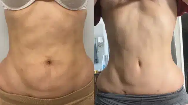
Self-Limiting Cases
Most cases of fat necrosis are self-limiting. They often resolve without any medical intervention. The body can absorb the necrotic tissue over time. This process may take several weeks to months. Patients usually experience no significant complications during this period.
Monitoring is essential in these situations. Regular check-ups help ensure that the condition does not worsen. Patients should report any changes in symptoms. This includes increased pain or swelling in the affected area.
Conservative Treatments
Conservative treatments focus on symptom management. These methods aim to relieve discomfort without invasive procedures. Common approaches include:
- Pain relief medications
- Warm compresses to reduce swelling
- Physical therapy for mobility issues
Pain relief medications, such as acetaminophen or ibuprofen, can be effective. They help manage pain and inflammation. Warm compresses can soothe the affected area. Physical therapy may be beneficial if mobility is impacted.
Lifestyle changes also play a role. Maintaining a healthy diet and staying active can support recovery. Patients should avoid activities that may exacerbate their symptoms.
Surgical Intervention
In severe cases, surgical intervention may become necessary. Surgery is typically considered when conservative treatments fail to provide relief. It may also be needed if there is a risk of complications.
Surgical options include:
- Excision of necrotic tissue
- Drainage of cysts or abscesses
Excision involves removing the damaged fat tissue entirely. This procedure can help alleviate pain and prevent further issues. Drainage may be required if fluid accumulates in the area, causing discomfort.
Radiation therapy is not commonly used for fat necrosis management. However, it may be applied in specific cases where other treatments are ineffective.
Follow-Up Care
Follow-up care is crucial after treatment. Regular evaluations allow healthcare providers to monitor progress. They can assess whether symptoms improve or require further intervention.
Patients should communicate openly with their healthcare team. Discussing concerns or changes in symptoms ensures timely adjustments to the treatment plan.
Pain Relief Strategies
Over-the-Counter Medications
Ibuprofen and acetaminophen are common over-the-counter pain relievers. These medications can help reduce discomfort associated with fat necrosis. Ibuprofen works as an anti-inflammatory, which can ease breast inflammation. Acetaminophen is effective for general pain relief. Both options are easily accessible and can provide quick relief for mild to moderate pain.
Dosage is important. Always follow the instructions on the label. If unsure, consult a healthcare provider for guidance. Regular use of these medications may be necessary during flare-ups. However, prolonged use should be avoided without medical advice.
Warm Compresses
Applying warm compresses can also alleviate pain. Heat encourages blood flow to the area and promotes healing. A simple method involves soaking a clean cloth in warm water. Wring it out and place it on the affected area for 15-20 minutes.
Warm compresses can be repeated several times a day. This method is safe and easy to do at home. It provides comfort without the side effects of medication. Many patients find this approach soothing during painful episodes.
Prescription Pain Medications
Consulting a healthcare provider is essential if over-the-counter options are insufficient. A doctor may prescribe stronger pain medications tailored to individual needs. Prescription medications can offer more effective relief for severe pain.
Healthcare providers assess each case carefully before prescribing medication. They consider factors like medical history and current health conditions. Communication about pain levels and symptoms helps them make informed decisions.
Medical professionals may also suggest alternative therapies alongside medications. These could include physical therapy or other non-invasive treatments. Such strategies can enhance overall management of fat necrosis.
Therapy Options
Therapy may play a role in managing discomfort from fat necrosis. Physical therapy focuses on exercises that improve mobility and reduce stiffness in the affected area. A trained therapist can design a program that fits individual needs.
e patients benefit from massage therapy as well. Gentle massage can relieve tension and promote relaxation in surrounding tissues. Always discuss any new therapies with a healthcare provider first.
Surgical Interventions
Surgery Necessity
Surgery may be necessary for managing fat necrosis. Large or painful lumps often require removal. These lumps can cause discomfort and affect quality of life. If the lumps are significant, doctors will recommend surgical intervention. This procedure aims to alleviate pain and restore normal function.
Malignancy Concerns
Surgical removal is also considered if there is a suspicion of malignancy. Doctors perform biopsies to examine tissue samples. This helps determine if cancer is present. If malignancy is suspected, swift action is critical. Early detection improves treatment outcomes significantly. Patients must discuss all concerns with their healthcare providers.
Imaging Studies
Additional imaging studies may be needed before surgery. This is especially true after breast surgery. Imaging helps assess the extent of changes in the tissue. Techniques like ultrasound or MRI can provide detailed views. These studies guide the surgical approach and ensure accurate procedures.
Procedure Details
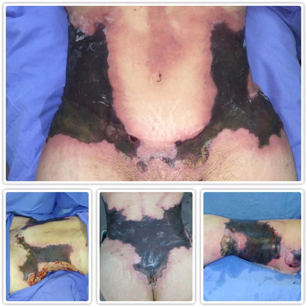
Specific procedures vary based on individual cases. Surgeons often perform significant tissue debridement during surgery. This involves removing damaged or dead tissue from the area. The goal is to promote healing and prevent complications.
Fat grafts are sometimes used in conjunction with these procedures. These grafts help fill defects left by surgery. They can improve the appearance of the breasts or buttocks after fat necrosis management.
Trauma Impact
Breast trauma plays a role in the development of fat necrosis. Understanding this connection is vital for effective management. Patients with a history of breast trauma should inform their doctors. This information helps tailor treatment plans.
In some cases, trauma may lead to significant changes in breast tissue. Surgical intervention can address these issues effectively. Doctors evaluate each case carefully before recommending surgery.
Recovery Considerations
Recovery from surgical interventions varies by individual and procedure type. Patients should follow post-operative care instructions closely. This includes managing pain, monitoring for infection, and attending follow-up appointments.
Regular check-ups allow doctors to assess healing progress. They can also identify any complications early on. Engaging in open communication with healthcare providers enhances recovery outcomes.
Potential Complications and Prognosis
Good Prognosis
Most cases of fat necrosis have a good prognosis. Many patients experience resolution without long-term issues. Symptoms often improve over time. Fat necrosis typically resolves itself, especially with proper management.
In some instances, patients may notice lumps in the affected area. These lumps can be alarming but are often harmless. Regular monitoring is crucial to ensure they do not signify underlying problems.
Possible Complications
e complications can arise from fat necrosis. Persistent pain is one of the most common issues. Patients might feel discomfort that lasts longer than expected. This pain can affect daily activities and quality of life.
Infections can also occur after fat grafting procedures. They may lead to further complications if not addressed promptly. Signs of infection include redness, swelling, and increased warmth around the area. If these symptoms appear, immediate medical attention is necessary.
Importance of Early Detection
Early detection plays a vital role in managing fat necrosis effectively. Recognizing symptoms early helps prevent complications. Patients should stay vigilant for any changes in their condition.
Appropriate management is essential to alleviate symptoms. Healthcare providers often recommend monitoring the affected area regularly. They may suggest imaging studies to assess the situation accurately. These steps help ensure that any potential issues are identified quickly.
For patients undergoing surgical interventions, understanding these aspects is crucial. Being aware of potential complications allows individuals to make informed decisions about their health.
Managing Symptoms
Managing symptoms effectively can lead to better outcomes. Pain relief options include medications and physical therapy. These treatments help reduce discomfort and improve mobility.
In some cases, additional procedures may be necessary to remove persistent lumps or tissue causing issues. Consulting with a healthcare provider ensures that the right course of action is taken.
Patients should discuss their concerns openly with their medical team. This communication fosters a supportive environment for recovery. It also allows for tailored treatment plans based on individual needs.
Importance of Patient Education
Postoperative Care
Educating patients about postoperative care is crucial. Proper care can significantly reduce the risk of fat necrosis. Patients should understand the importance of following their surgeon’s instructions closely. This includes taking prescribed medications and avoiding certain activities for a specified time.
Patients must also learn how to care for their surgical sites. Keeping the area clean and dry helps prevent infections, which can increase the likelihood of complications. Surgeons should provide clear guidance on wound care and hygiene practices.
Signs and Symptoms
Informing patients about the signs and symptoms of fat necrosis is essential. Patients need to know what to watch for after surgery. Common signs include lumps in the tissue, swelling, and changes in skin color. Recognizing these symptoms early can lead to prompt treatment.
Patients should be encouraged to report any unusual changes immediately. Quick action can prevent further complications or more severe issues. Educating them about these warning signs empowers them to take charge of their recovery.
Regular Follow-Ups
Regular follow-ups are vital, especially for high-risk individuals. Patients with a history of fat necrosis or other complications may need closer monitoring. These appointments allow healthcare providers to assess healing and address any concerns.
During follow-ups, doctors can evaluate the surgical site and check for any signs of necrosis. They can also adjust treatment plans based on individual needs. This ongoing communication fosters a supportive environment for recovery.
Patients should be reminded to schedule follow-up visits as directed by their healthcare team. Consistent monitoring ensures that any emerging issues are caught early.
High-Risk Individuals
Certain groups may be at higher risk for developing fat necrosis. For example, individuals with obesity or those who smoke may face increased risks after surgery. Educating these patients on their specific risks is important.
They should receive tailored advice on lifestyle changes that could aid recovery. Discussing nutrition, exercise, and smoking cessation can help improve outcomes. Encouragement from healthcare providers can motivate patients to make healthier choices.
Emotional Support
Providing emotional support is also part of patient education. Recovery from surgery can be stressful and overwhelming. Patients may experience anxiety about complications like fat necrosis.
Offering resources such as counseling or support groups can help alleviate fears. Patients who feel supported are more likely to engage in their recovery actively.
Final Remarks
Fat necrosis can be a challenging condition to manage, but understanding its causes, symptoms, and treatment options is crucial. You’ve explored various aspects of fat necrosis, from diagnosis to pain relief strategies. Each section highlights the importance of proactive management and patient education.
Stay informed about your health and seek timely medical advice if you suspect fat necrosis. Knowledge empowers you to make better decisions for your well-being. Don’t hesitate to discuss concerns with your healthcare provider. Together, you can navigate this condition effectively and improve your quality of life.
Frequently Asked Questions
What is fat necrosis?
Fat necrosis is a condition characterized by the death of fat tissue, often resulting in lumps or nodules. It can occur due to trauma, surgery, or inflammation.
What causes fat necrosis?
Fat necrosis is commonly caused by physical trauma, surgical procedures, infections, or certain medical conditions like pancreatitis.
Who is at risk for developing fat necrosis?
Individuals who have undergone surgery, especially breast surgery, are at higher risk. Those with conditions that cause fatty tissue inflammation may also be affected.
How is fat necrosis diagnosed?
Diagnosis typically involves a physical examination and imaging tests such as ultrasound or MRI. A biopsy may be performed if the diagnosis is uncertain.
What are the treatment options for fat necrosis?
Management includes observation for asymptomatic cases and pain relief strategies. Surgical removal may be necessary for larger or painful lesions.
Can fat necrosis lead to complications?
While generally not serious, complications can include persistent pain or infection. Most cases resolve without significant issues.
How important is patient education in managing fat necrosis?
Patient education is crucial for understanding symptoms, treatment options, and when to seek medical advice. Informed patients can better manage their condition effectively.